
Why Should I Get a Glaucoma Eye Exam?
What if I told you that a simple glaucoma eye exam could be the key to preserving your vision against a condition known as the “silent thief”? There’s a common misconception that if you have perfect vision, you’re safe from glaucoma, but perfect vision doesn’t guard against the internal eye pressure changes that glaucoma brings.
I’m Minh Van Tran, owner and principal optometrist of VisionPro in Footscray and St. Albans. I’ve been taking care of patients here since 2008, with particular focus on helping our elderly community maintain good vision as they age. With a long background in optometry and a keen interest in managing ocular diseases, I aim to provide care that’s both understanding and thorough. At VisionPro, we believe in keeping you informed and comfortable with your eye care choices.
Early and regular eye exams can significantly reduce the risk of vision loss which glaucoma brings, allowing you to enjoy life’s moments fully.
Why risk your eyesight? Book your appointment now and ensure your eyes receive professional attention. Or keep exploring this article to understand more about how our personalized eye care can make a difference.
Understanding Glaucoma: The Silent Thief of Sight
Glaucoma is a progressive eye condition that can lead to irreversible vision loss. It primarily affects the optic nerve, responsible for transmitting visual information from the eye to the brain. The damage often stems from increased intraocular pressure (IOP), due to a build-up of aqueous humor, a fluid that flows through the eye.
- Open-angle glaucoma: This is the most common type, developing gradually as the eye’s drainage channels become inefficient. This leads to a build-up of aqueous humor, increasing IOP. The optic nerve is gradually damaged, causing a slow loss of peripheral vision. This progression often goes unnoticed until significant vision loss has occurred.
- Angle-closure glaucoma: This form occurs suddenly when the drainage angle between the iris and cornea closes, causing a rapid increase in IOP. This often leads to immediate symptoms such as eye pain, blurred vision, and headaches. Angle-closure glaucoma requires urgent medical attention to prevent permanent damage to the optic nerve.
Identifying the symptoms of glaucoma is essential for timely intervention and treatment, which are critical to preventing the potentially severe consequences of this condition.

Risk Factors for Glaucoma
It is crucial to understand the risk factors for glaucoma, if we aim for early detection and effective management of this vision-threatening condition.
- Age: The risk of glaucoma increases significantly for individuals over 60. Regular eye exams become crucial to monitor eye health and detect any early signs of glaucoma.
- Family history: A genetic predisposition can increase the likelihood of developing glaucoma. Individuals with a family history of the condition should undergo regular check-ups to detect any early signs.
- Diabetes: Individuals with diabetes are at a heightened risk for developing glaucoma due to potential changes in blood vessels that affect the retina and the optic nerve. Managing blood sugar levels and undergoing regular eye exams are critical.
- High Blood Pressure: Circulation of blood to the optic nerve, can be impaired by high blood pressure, potentially reducing the vital supply of nutrients and oxygen, which increases the risk of damage.
- High Cholesterol: Elevated cholesterol levels can lead to vascular complications that affect ocular health, potentially impacting the optic nerve due to restricted blood flow.
By staying informed about these risk factors and maintaining regular eye health screenings, patients can enhance their chances of preventing or managing glaucoma effectively.
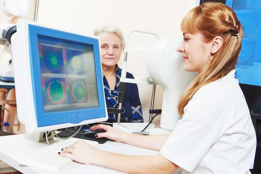
Eye Exams for Glaucoma
Here’s a detailed look at glaucoma eye exams which we might perform at our clinic to examine your eye health condition.
| Test Type | Description |
|---|---|
| Puff of Air Tonometry | Measures eye pressure through a quick puff of air, aimed at your eye. Known as non-contact tonometry, it temporarily flattens your cornea to test resistance to the air puff. This calculates and measures the intraocular pressure. It provides a useful preliminary measurement but is less precise than other methods. |
| Goldman Tonometer Test | Considered the gold standard in tonometry, this test uses a small probe that touches the cornea to measure internal eye pressure. Highly accurate and typically conducted after numbing the eye, it measures the force needed to flatten part of the cornea, translating this into a pressure reading. |
| Ophthalmoscopy | Allows an optometrist to view the inside of the back of your eye, examining the optic nerve, crucial for glaucoma diagnosis. The optometrist uses a hand-held device with a light to inspect the optic nerve’s color and shape. Unusual optic nerve appearance or a high cup-to-disc ratio may indicate glaucoma. |
| Perimetry | Known as a visual field test, it examines all areas of eyesight, especially peripheral vision, which glaucoma often affects first. You indicate when a moving object enters your peripheral vision, helping map visual fields and identify patterns of vision loss. |
| Dilated Eye Exams | Involves using drops to dilate the pupils, allowing a special magnifying lens to examine the retina and optic nerve more clearly. This exam provides crucial information about these structures’ health. |
| Optic Nerve Photography | Captures detailed images of the optic nerve with a special photographic device. These images are valuable for documenting the nerve’s current state and can serve as a baseline for comparing future changes, assisting in monitoring glaucoma progression. |
| Optical Coherence Tomography | OCT is a sophisticated scanning system that produces a cross-sectional image of the retina. It measures the retina’s and optic nerve fibers’ thickness, providing detailed structural information vital for early glaucoma detection. |
| Pachymetry | Measures the thickness of the cornea, the clear front layer of the eye. Since corneal thickness can influence eye pressure readings, knowing this can help interpret tonometry results more accurately. |
| Gonioscopy | Inspects the eye’s drainage angle—where eye fluids drain out—to ascertain if the angle is open, narrow, or closed. This helps determine the glaucoma type and guide treatment options. |
| Corneal Hysteresis | Measures the cornea’s ability to absorb and dissipate energy, reflecting its biomechanical properties. Lower corneal hysteresis is associated with a higher risk of glaucoma progression, indicating the importance of this measure in glaucoma risk assessment. |
| Visual Evoked Potential (VEP) | Measures the electrical activity in the brain’s vision center to check if visual information is being transmitted properly from the eye to the brain. This test is crucial for identifying issues in advanced glaucoma cases. |
| Electroretinogram (ERG) | Assesses the electrical responses of the eye’s light-sensitive cells (rods and cones). These responses help determine if these cells are healthy and processing light correctly, which can be compromised in glaucoma. |
If you’d like to learn more about glaucoma eye exams, watch the following video by Dr Joseph Allen as he explains the variety of ways that optometrists and eye doctors can test for the condition.
Treatment Options
Fortunately, there are several treatment options available that aim to lower intraocular pressure (IOP), which is crucial in slowing the progression of the disease. Here’s a closer look at the typical treatments:
Medications
Medications are often the first line of defense against glaucoma. These usually come in the form of eye drops but can also include oral medications, especially in more severe cases. The primary goal of these medications is to lower the eye pressure either by reducing the amount of aqueous humor (the fluid inside the eye) the eye produces or by improving the flow through the drainage angle, helping the fluid to drain out of the eye more efficiently.
- Eye Drops: There are several types of eye drops used to treat glaucoma, each working in a different way to help lower eye pressure. For example, Prostaglandin analogs increase the outflow of the fluid in your eye and are often prescribed because of their effectiveness and the convenience of needing only once-daily dosing. Beta-blockers, which reduce the production of fluid, are also commonly used but might also have a variety of side effects.
- Oral Medications: Sometimes, when eye drops are not sufficient to bring down the eye pressure, oral medications may be prescribed. These are usually carbonic anhydrase inhibitors, which reduce the production of aqueous humor. Oral medications are generally used for short periods because they can have more systemic side effects compared to eye drops.
Laser Therapy
Laser treatment offers a middle ground between medications and more invasive surgery and can be used for both open-angle and angle-closure glaucoma:
- Trabeculoplasty: This is a common laser treatment for open-angle glaucoma, the most common form of the disease. The laser is used to open clogged drainage canals in the trabecular meshwork (the eye’s drainage system), aiming to improve the outflow of aqueous humor from the eye and thereby reduce intraocular pressure.
- Iridotomy: This is often used for angle-closure glaucoma. In this procedure, the laser creates a small hole in the iris, helping to open up the angle that drains the fluid from the eye, which can prevent or alleviate sudden increases in eye pressure.
Eye Surgery
In cases where medications and laser therapy do not adequately control eye pressure, or where the glaucoma is progressing rapidly, more invasive surgeries may be necessary:
- Trabeculectomy: This is a common surgical procedure for glaucoma where a new drainage pathway for aqueous humor is created to bypass the blocked or malfunctioning trabecular meshwork. By allowing fluid to drain from the eye through this new pathway, eye pressure is reduced.
- Glaucoma Implant Surgery: Another option is to implant a tiny drainage device in the eye, which helps to increase the outflow of fluid and lower eye pressure. This device creates a new pathway for fluid to exit the eye, helping to maintain a healthier level of intraocular pressure.
- Minimally Invasive Glaucoma Surgeries (MIGS): These procedures are newer and generally involve less risk than traditional glaucoma surgeries. MIGS typically have shorter recovery times and can be an effective option for patients with mild-to-moderate glaucoma.
Each of these treatments has its own set of potential benefits and risks, and the best approach depends on the specific type and severity of glaucoma, as well as the patient’s overall health and personal circumstances. Managing glaucoma is a lifelong process that requires regular monitoring and treatment adjustments to preserve vision as much as possible. Having regular glaucoma eye exams and working closely with a healthcare provider to customize treatment can provide the best defense against the serious consequences of this eye disease.
VisionPro and Glaucoma Eye Exams
At VisionPro we emphasize the importance of ongoing care. We support our patients in managing glaucoma through regular check-ups and co-management with their ophthalmologist, if they have one. We also guide our patients on making important lifestyle adjustments to lower their glaucoma risk factors or slow glaucoma progression. Some suggestions include:
- Diet: Consuming foods rich in vitamins and antioxidants, such as leafy greens, berries, and fish, can contribute to eye health. These nutrients can help support overall well-being and potentially reduce the risk of glaucoma progression.
- Exercise: Regular physical activity can improve overall well-being and may help lower intraocular pressure, reducing strain on the optic nerve. Exercise also supports cardiovascular health, which can indirectly benefit eye health.
- Avoiding smoking and alcohol: Smoking has been linked to various eye conditions, including glaucoma, and can exacerbate its progression. Limiting alcohol consumption is also important, as excessive intake can raise intraocular pressure, further damaging the optic nerve.
By making these changes and adhering to regular glaucoma eye exams, individuals can manage the condition effectively, preserving their vision and quality of life.
Final Thoughts
Glaucoma, often undetected until significant vision loss occurs, necessitates regular eye exams for early intervention. Failing to act while there is still time could result in a preventable deterioration of your eye health and vision quality.
Don’t wait for a sign that might never come; be proactive about your eye health.
Scheduling an eye exam appointment is the first step to restoring or maintaining quality of life.
You can call us on (03) 9687 8787 for Footscray, (03) 9364 5509 for St Albans, (03) 9600 1142 for Sunshine OR arrange an appointment using the “Book An Appointment” button in the navigation menu. All our practices are easily accessible, with nearby parking and public transport options available.

Minh gained his Bachelor of Optometry in 2000, and his Certificate in Ocular Therapeutics (ACO) in 2016.
He started VisionPro Optometrists in 2008 and has been Principal Optometrist ever since, working in both the Footscray and the St Albans practices.
Minh is a member of Optometrist Association Australia and the ACO. He always strives to achieve the highest standard of professionalism when delivering eyecare to all his patients.
Minh’s special areas of interest include ocular diseases and management, children’s vision and contact lens fitting. Minh enjoys travelling and reading in his spare time.